Keratoconus Diagnosis in Adelaide

Blurred vision, distorted vision, glare, halos, and frequent prescription changes can be signs of keratoconus.

At Innovative Eye Care, we provide advanced keratoconus diagnosis in Adelaide and Henley Beach using detailed corneal imaging and evidence-based clinical assessment. Our optometrists assess the shape, thickness, strength, and optical quality of your cornea to detect keratoconus early and monitor changes over time.

Book a keratoconus assessment
Innovative Eye Care clinic in Adelaide for keratoconus diagnosis and management

What is Keratoconus?

Keratoconus is a progressive condition that affects the cornea, the clear front surface of the eye. In keratoconus, the cornea becomes thinner and more irregular. It can bulge into a cone-like shape, which makes light focus unevenly inside the eye. This can cause blurred, distorted vision that may not improve fully with standard glasses.1–3

Keratoconus often starts during the teenage years or early adulthood. It can continue changing into the 30s or 40s, so early diagnosis and regular monitoring matter.1,2,4,5

Learn more about keratoconus treatment and management
Innovative Eye Care clinic in Adelaide for keratoconus diagnosis and management

Signs You May Need a Keratoconus Assessment

Book an assessment if you notice any of the following.6,7

  • Blurred or distorted vision
  • Glare or halos, especially at night
  • Increasing light sensitivity
  • Frequent prescription changes
  • Increasing astigmatism
  • Poor vision even with updated glasses
  • One eye seeing worse than the other
  • A family history of keratoconus
  • A history of eye rubbing, allergies, hay fever, asthma, or eczema

Early keratoconus can be hard to detect with a standard eye test alone. Advanced corneal imaging gives your optometrist a much clearer view of subtle changes in the cornea.2,7,8

Pentacam corneal tomography maps used for keratoconus diagnosis

How Keratoconus is Diagnosed

Keratoconus diagnosis starts with a detailed eye examination and corneal imaging.9,10

At Innovative Eye Care, your assessment may include:
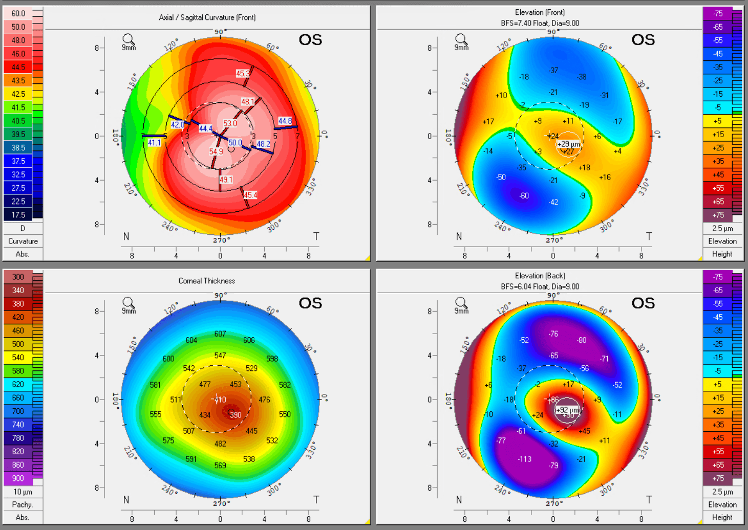
Corneal tomography

This creates a 3D map of the cornea, including the front and back surfaces. It helps detect early keratoconus and measure corneal thickness.5,9,11,12
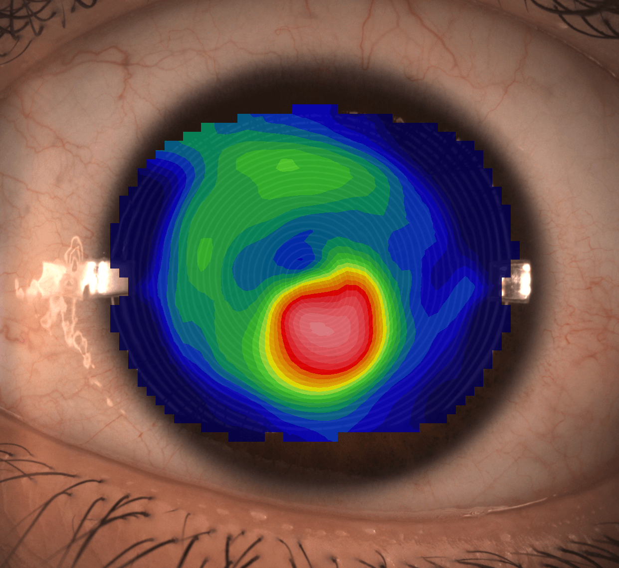
Corneal topography map showing keratoconus on the eye

Corneal topography

This maps the front surface of the cornea. It helps identify irregular astigmatism and corneal steepening.9
Learn more about corneal topography
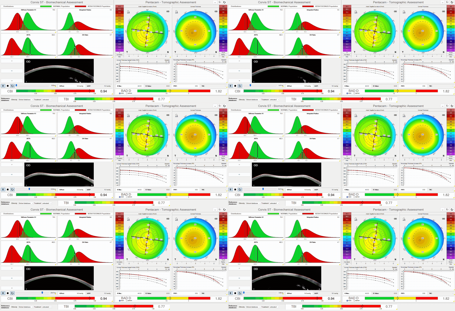
Pentacam and Corvis ST assessment for keratoconus diagnosis and monitoring

Corneal biomechanics testing

This measures how the cornea responds to pressure. It can help identify corneas that may be structurally weaker.10,11,13
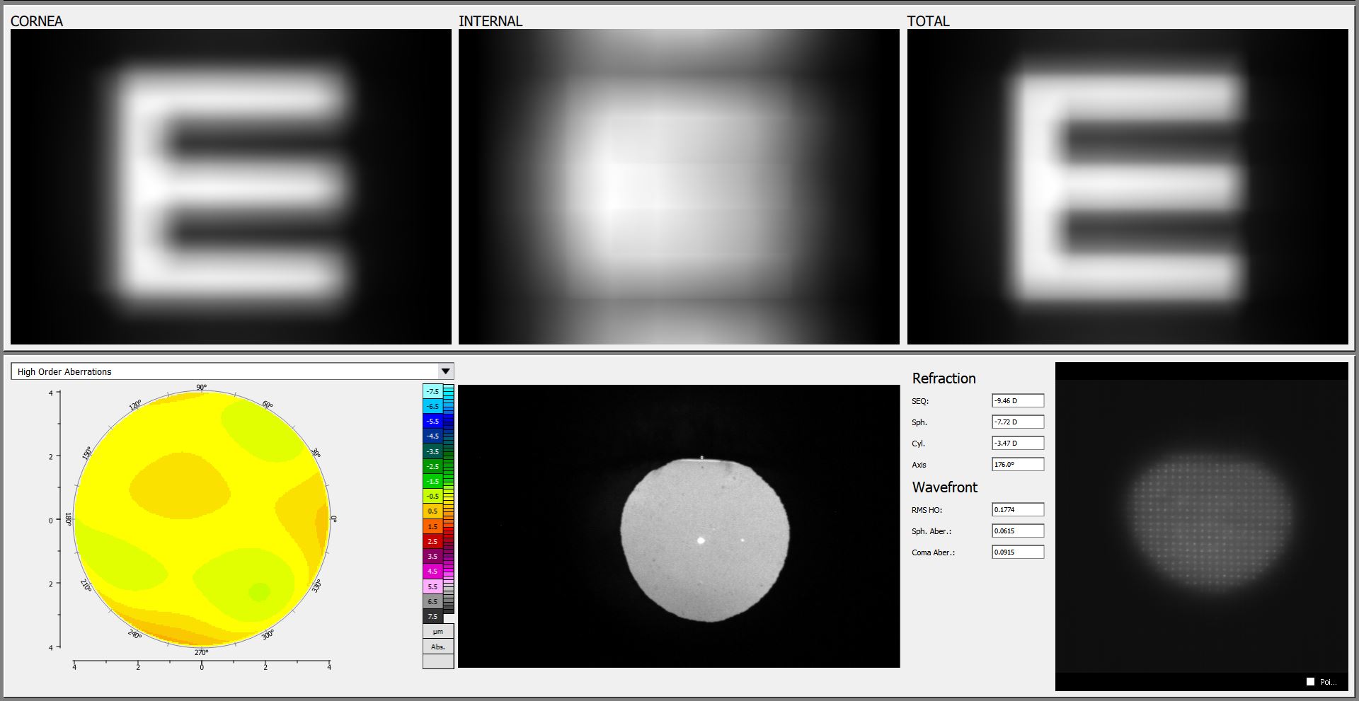
Wavefront aberrometry scan used in keratoconus diagnosis at Innovative Eye Care Adelaide

Wavefront Aberrometry

This measures higher-order optical distortions that can cause glare, halos, ghosting, and reduced clarity.7
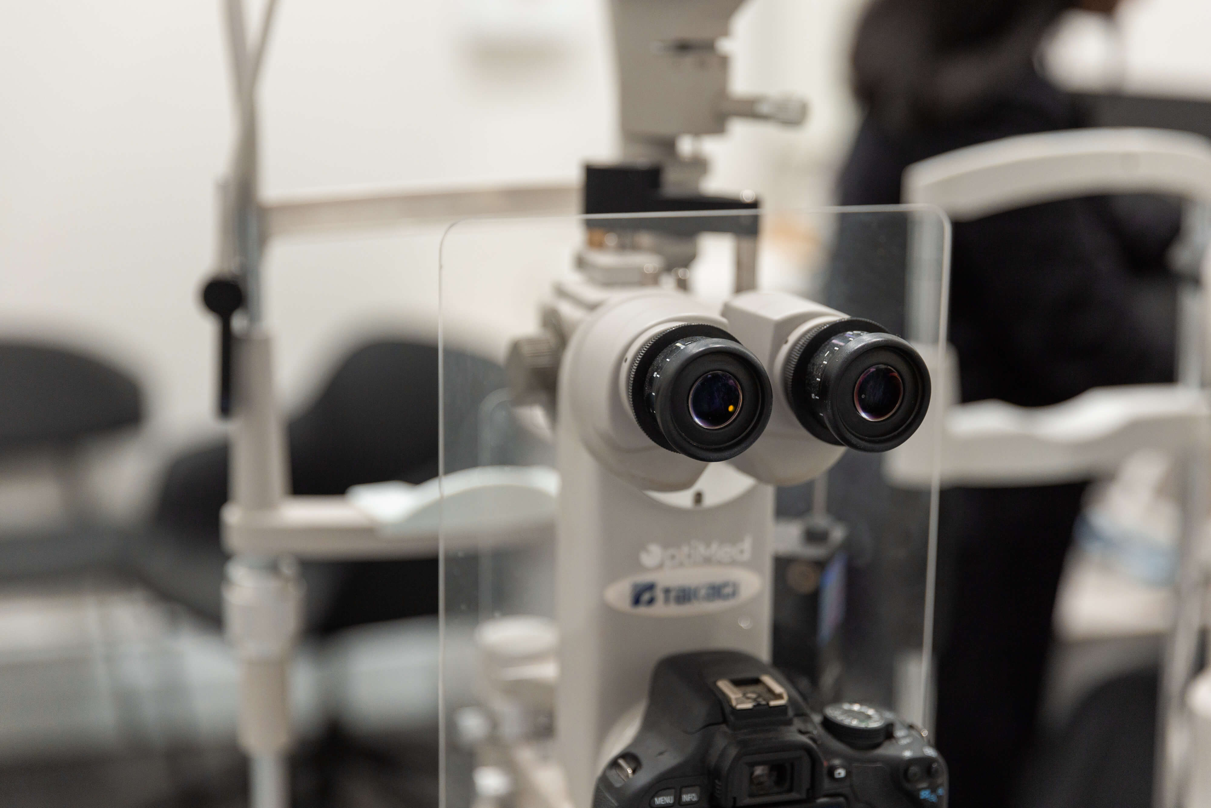
Slit lamp microscope used for keratoconus diagnosis at Innovative Eye Care Henley Beach

Slit lamp examination

This allows your optometrist to assess the corneal surface and look for clinical signs of keratoconus.
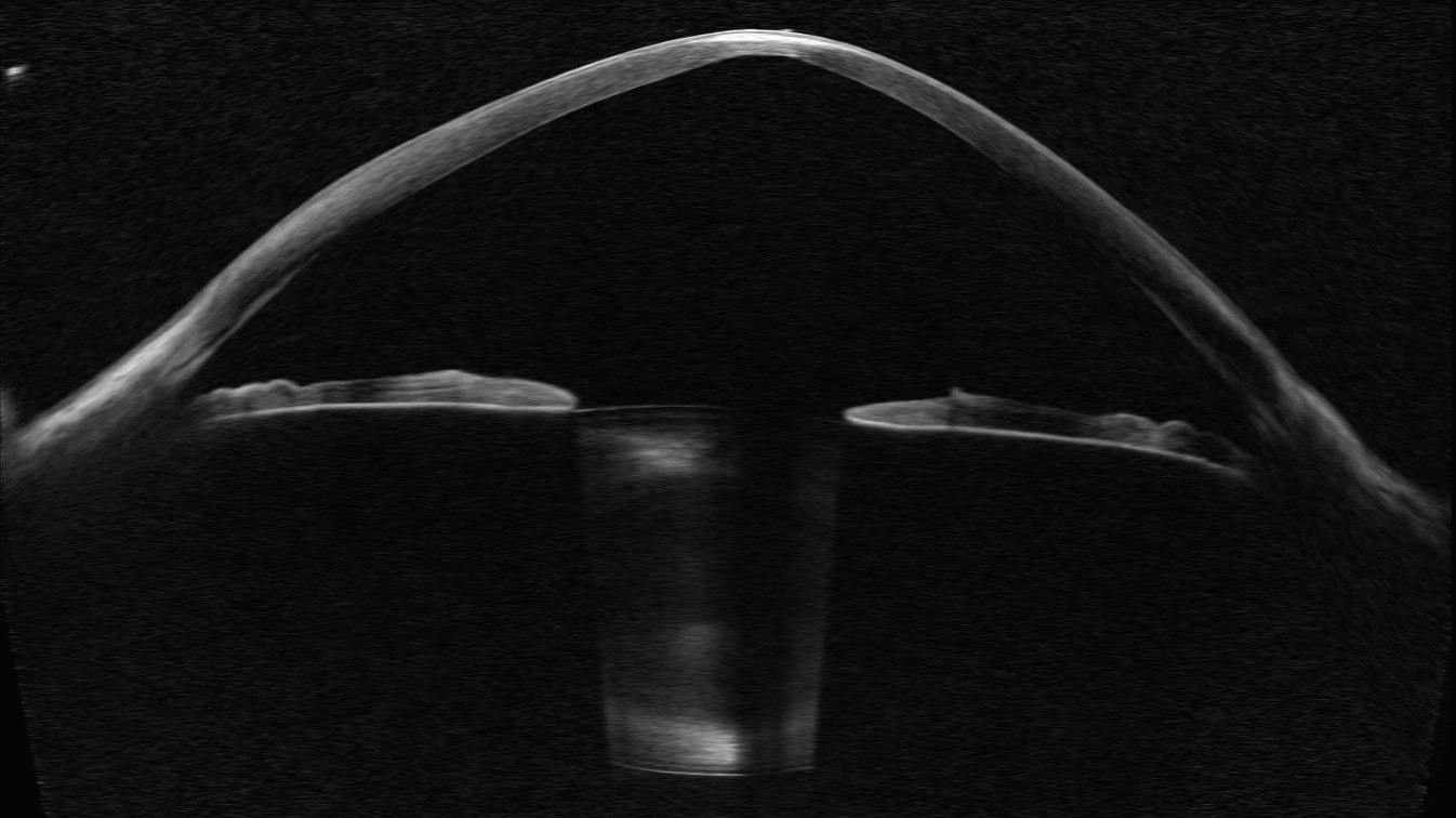
Anterior OCT

This provides cross-sectional imaging of the cornea and can help assess corneal shape, thickness, and contact lens fitting.
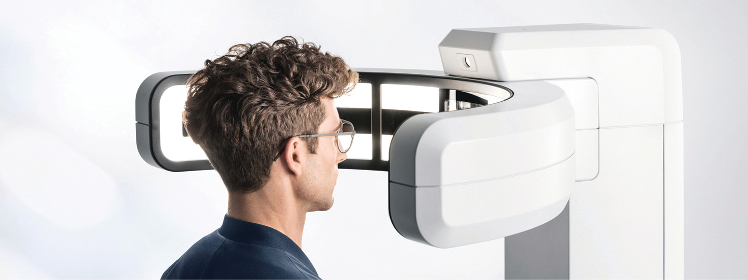
Innovative Eye Care clinic in Adelaide for keratoconus diagnosis and management

Why Early Keratoconus Diagnosis Matters

Early diagnosis gives you more options.

Keratoconus can progress before symptoms feel severe.7,10,14 By the time vision feels noticeably distorted, the cornea may already have changed significantly.

A diagnosis helps your optometrist:

  • Confirm whether keratoconus is present
  • Measure the severity of the condition
  • Track whether it is progressing
  • Recommend the right vision correction
  • Refer for corneal collagen cross-linking when needed
  • Reduce avoidable risks, such as eye rubbing
  • Plan specialty contact lenses if glasses no longer provide clear vision

Corneal collagen cross-linking is most effective when progressive keratoconus is detected early.5,14 Your optometrist can refer you to an ophthalmologist if your results suggest progression or higher risk.
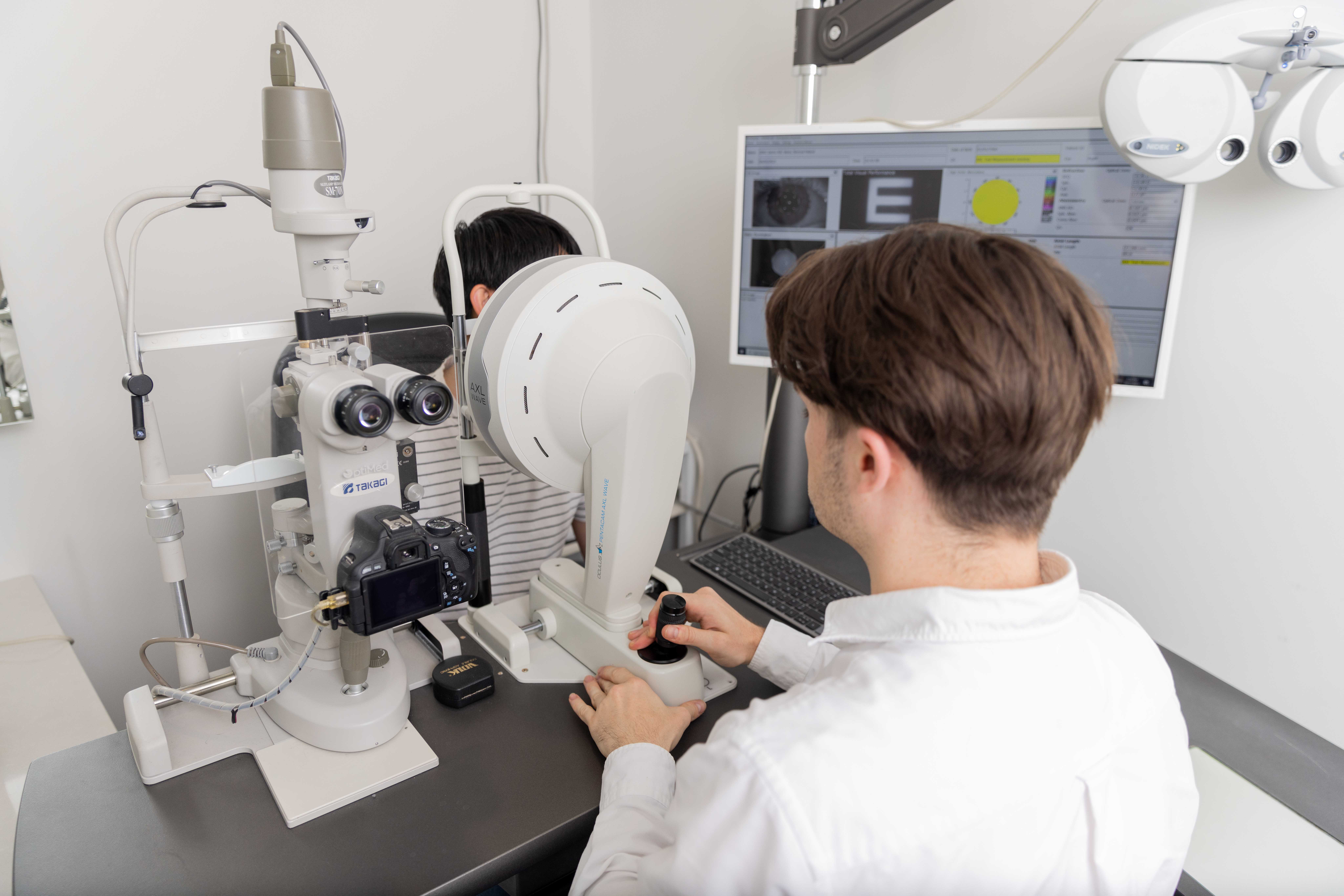
Optometrist performing corneal scans for keratoconus diagnosis in Adelaide

What Happens During a Keratoconus Assessment?

Your keratoconus assessment is designed to understand both your vision and your corneal health.

Your visit may include:

  • A review of your symptoms and eye history
  • Questions about allergies, eye rubbing, and family history
  • Vision testing
  • Refraction to measure your prescription
  • Corneal topography and tomography
  • OCT imaging where required
  • Assessment of glare, halos, and visual distortion
  • Contact lens suitability discussion if needed
  • A clear management plan

You will be told whether your results suggest keratoconus, whether further monitoring is needed, and what the next step should be.
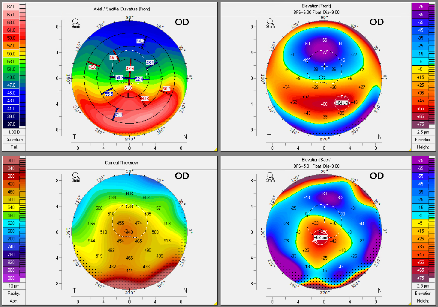
Corneal tomography showing PMD with steepening and thinning

Keratoconus and Specialty Contact Lenses

In early keratoconus, glasses or soft contact lenses may still provide useful vision.

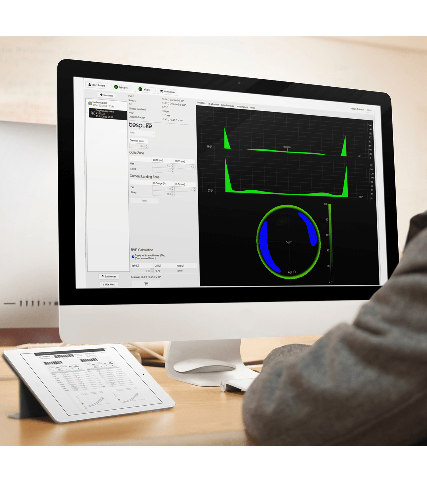
As the cornea becomes more irregular, glasses often become less effective. Specialty contact lenses can help by creating a smoother optical surface over the cornea.

Options may include.5,15

  • Rigid gas permeable lenses
  • Scleral contact lenses
  • Custom soft lenses
  • Hybrid lens designs in selected cases

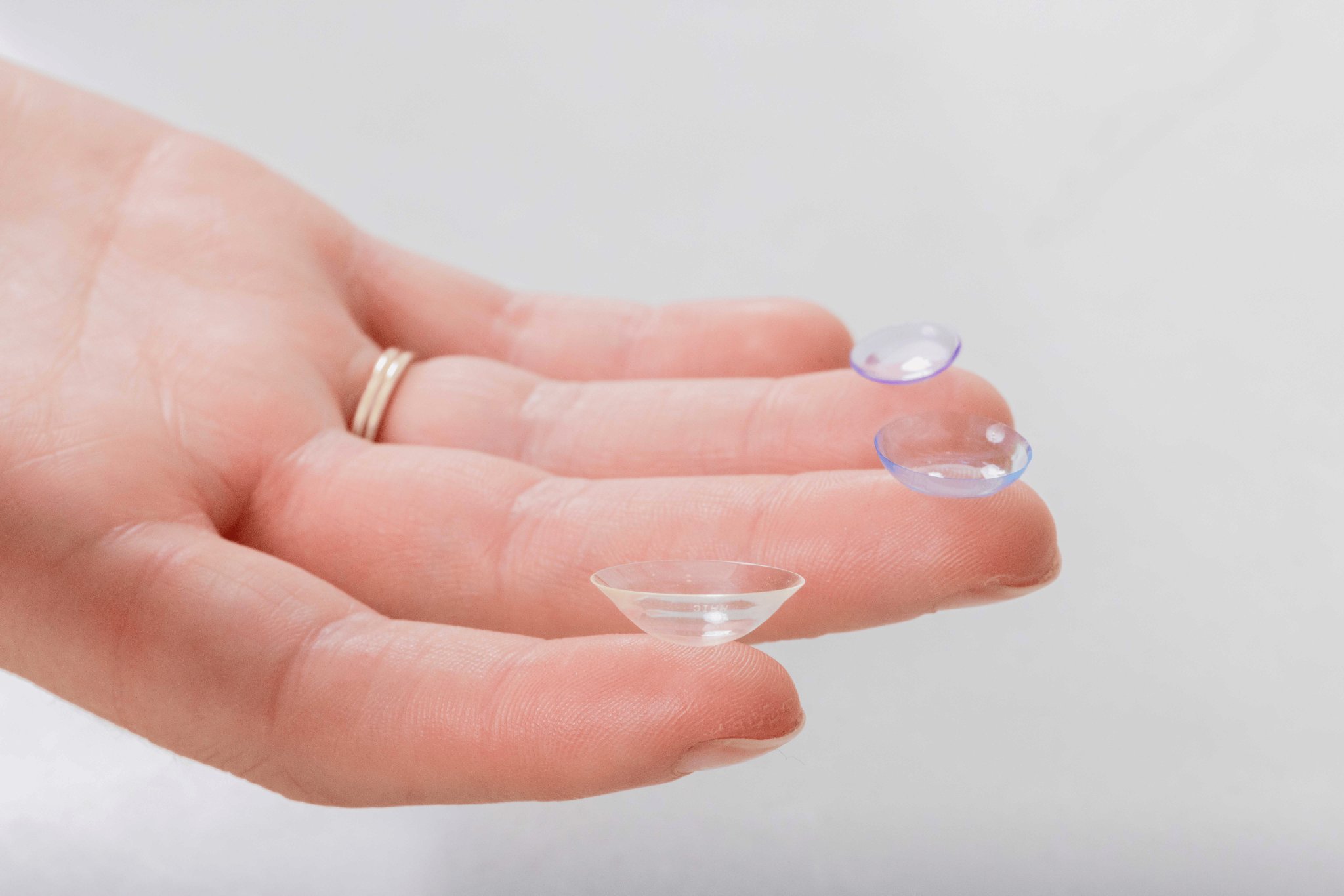
Scleral lenses are often used for moderate to advanced keratoconus.16 They vault over the cornea and rest on the white part of the eye, which can improve comfort and vision for irregular corneas.
Learn more about scleral contact lensesLearn more about contact lensesLearn more about contact lens fitting and costs
Corneal tomography showing PMD with steepening and thinning

Keratoconus vs Pellucid Marginal Degeneration

Pellucid marginal degeneration, or PMD, is another corneal thinning condition that can look similar to keratoconus. PMD usually affects the lower cornea and can cause high against-the-rule astigmatism.17

Accurate diagnosis is important because the contact lens fitting and management plan may differ. Advanced corneal imaging helps your optometrist distinguish between these conditions.

Innovative Eye Care clinic in Adelaide for keratoconus diagnosis and management

Why choose Innovative Eye Care for Keratoconus Diagnosis?

Keratoconus diagnosis and management is a core clinical focus at Innovative Eye Care.

Our team uses advanced imaging technology to assess corneal shape, thickness, biomechanics, and optical quality. We also have extensive experience fitting specialty contact lenses for keratoconus and other irregular corneal conditions.

Principal optometrist Lachlan Hoy has written a textbook chapter on keratoconus and regularly contributes to education in specialty contact lens practice.
Meet our optometrists
Innovative Eye Care clinic in Adelaide for keratoconus diagnosis and management

Book a Keratoconus Assessment in Adelaide or Henley Beach

If you have blurred vision, increasing astigmatism, frequent prescription changes, night glare, or a family history of keratoconus, book a keratoconus assessment.

Innovative Eye Care provides keratoconus diagnosis and monitoring at our Adelaide and Henley Beach practices.

Book or contact usView our Adelaide and Henley Beach locations:

Keratoconus Diagnosis FAQs

Can an optometrist diagnose keratoconus?
Yes. An optometrist with the right diagnostic technology can detect and monitor keratoconus. Advanced imaging such as corneal tomography and topography is important because early keratoconus may not be obvious during a standard eye test.
What is the best test for keratoconus?
Corneal tomography is one of the most important tests for keratoconus because it maps the front and back surfaces of the cornea and measures corneal thickness. Your optometrist may also use topography, OCT, wavefront aberrometry, and corneal biomechanics testing.
What are the first signs of keratoconus?
Early signs can include blurred vision, ghosting, glare, halos, light sensitivity, increasing astigmatism, and frequent prescription changes. Some people notice that glasses no longer give sharp vision.
Is keratoconus serious?
Keratoconus can become serious if it progresses and causes significant corneal thinning or scarring. Early diagnosis and monitoring can help protect long-term vision and guide timely treatment.
Can keratoconus be stopped?
Corneal collagen cross-linking can slow or stop progression in suitable cases. Your optometrist can monitor your cornea and refer you to an ophthalmologist if cross-linking may be needed.
Do glasses fix keratoconus?
Glasses may help in early keratoconus. As the cornea becomes more irregular, glasses often become less effective. Specialty contact lenses, including rigid gas permeable or scleral lenses, may provide clearer vision.
Where can I get keratoconus diagnosis in Adelaide?
Innovative Eye Care provides keratoconus diagnosis and monitoring at Adelaide and Henley Beach, using advanced corneal imaging and specialty contact lens expertise.

References

  1. Rabinowitz YS. Keratoconus. Surv Ophthalmol. 1998;42(4):297-319. doi:10.1016/s0039-6257(97)00119-7
  2. Romero-Jiménez M, Santodomingo-Rubido J, Wolffsohn JS. Keratoconus: a review. Contact Lens Anterior Eye J Br Contact Lens Assoc. 2010;33(4):157-166; quiz 205. doi:10.1016/j.clae.2010.04.006
  3. Lawless M, Coster DJ, Phillips AJ, Loane M. Keratoconus: diagnosis and management. Aust N Z J Ophthalmol. 1989;17(1):33-60. https://pubmed.ncbi.nlm.nih.gov/2527524/
  4. Chan E, Chong EW, Lingham G, et al. Prevalence of Keratoconus Based on Scheimpflug Imaging: The Raine Study. Ophthalmology. 2021;128(4):515-521. doi:10.1016/j.ophtha.2020.08.020
  5. Singh R, Koh S, Sharma N, et al. Keratoconus. Nat Rev Primer. 2024;10(1):81. doi:10.1038/s41572-024-00565-3
  6. Hashemi H, Heydarian S, Hooshmand E, et al. The Prevalence and Risk Factors for Keratoconus: A Systematic Review and Meta-Analysis. Cornea. 2020;39(2):263-270. doi:10.1097/ICO.0000000000002150
  7. Santodomingo-Rubido J, Carracedo G, Suzaki A, Villa-Collar C, Vincent S. Keratoconus: An updated review. Cont Lens Anterior Eye. 2022;45(3):101559. doi:10.1016/j.clae.2021.101559
  8. Dina MS, Marinescu MC, Corbu CG, Constantin MM, Tataru CI, Tataru CP. Corneal Topography – a Review of Available Investigation Methods and Impact in the Diagnosis and Follow-Up of Keratoconus. Maedica - J Clin Med. 2025;20(2). doi:10.26574/maedica.2025.20.2.374
  9. Gomes JAP, Tan D, Rapuano CJ, et al. Global Consensus on Keratoconus and Ectatic Diseases. Cornea. 2015;34(4):359-369. doi:10.1097/ICO.0000000000000408
  10. Wang X, Maeno S, Wang Y, et al. Early diagnosis of keratoconus using corneal biomechanics and OCT derived technologies. Eye Vis. 2025;12(1):18. doi:10.1186/s40662-025-00435-3
  11. Bevara A, Vaddavalli PK. The Evolution of Diagnostics for Keratoconus: From Ophthalmometry to Biomechanics. Semin Ophthalmol. 2023;38(3):265-274. doi:10.1080/08820538.2022.2152716
  12. Kuo AN, Cortina MS, Greiner MA, et al. Advanced Corneal Imaging in Keratoconus. Ophthalmology. 2024;131(1):107-121. doi:10.1016/j.ophtha.2023.07.030
  13. Bao F, Geraghty B, Wang Q, Elsheikh A. Consideration of corneal biomechanics in the diagnosis and management of keratoconus: is it important? Eye Vis. 2016;3(1):18. doi:10.1186/s40662-016-0048-4
  14. Bui A, Truong A, Parischa N, Indaram M. Keratoconus Diagnosis and Treatment: Recent Advances and Future Directions. Clin Ophthalmol. 2023;17:2705-2718. doi:10.2147/OPTH.S392665
  15. Rathi M, Mandathara P, Dumpati S. Contact lens in keratoconus. Indian J Ophthalmol. 2013;61(8):410-415. doi:10.4103/0301-4738.116066
  16. Levit A, Benwell M, Evans B. Randomised controlled trial of corneal vs. scleral rigid gas permeable contact lenses for keratoconus and other ectatic corneal disorders. Cont Lens Anterior Eye. 2020;43(6):543-552. doi:10.1016/j.clae.2019.12.007
  17. Tsatsos M, Koulotsiou K, Giachos I, Tsinopoulos I, Ziakas N. Pellucid Marginal Degeneration: A Comprehensive Review of Pathophysiology, Diagnosis, and Management Strategies. J Clin Med. 2025;14(15):5178. doi:10.3390/jcm14155178

Speak to our friendly team today

Book your appointment now for personalised eye care tailored just for you.